
(Credit: Natasha Connell/Unsplash )
Brain MRIs may reveal psychotic disorder risk
Using MRI, researchers have found that people at risk of psychotic disorders have dysfunction in their brain's striatum.
Neurological markers in the brain can help detect people at risk of developing psychotic disorders and indicate when this risk has been successfully treated, researchers say.
People who may hear and see things that are not there could have symptoms of psychosis, better known as psychotic disorders.
“Psychotic disorders like schizophrenia are often lifelong and disabling for individuals,” says John Kerns, professor of psychology at the University of Missouri. “These disorders have major public health and societal costs greater than cancer. A major goal of our current research is to understand the nature of psychosis risk so we can prevent years of suffering.”
Researchers say psychotic disorders are associated with increased levels of dopamine—a chemical released by nerve cells—in a subregion of the brain called the striatum. This area is wired to process positive versus negative feedback for learning, often resulting in a person’s thoughts and actions based on what they’ve experienced in the past. Therefore, researchers suggest that these disorders involve a faulty feedback in learning that then drives a person’s faulty beliefs and perceptions. However, measuring levels of dopamine in people is costly, invasive, and not feasible in everyday clinical practice.
In this new study, researchers used an MRI and found that people at risk for the disorders exhibit evidence of dysfunction in the striatum.
“This dysfunction is most evident when performing tasks where people need to learn from positive and negative feedback,” Kerns says. “For instance, we have found that the risk for psychotic disorders involves increased activation in the striatum for positive feedback, and negative feedback involves decreased activation in the same subregion of the brain.”
Researchers believe this pattern of activation could explain symptoms of psychotic disorders. For example, activation resulting from increased positive feedback could make a person’s assumption seem truer than it really is, meanwhile activation from decreased negative feedback could make someone less likely to discard negative ideas.
The team will conduct future research to examine how well an MRI can predict the risk of psychotic disorders and whether prevention treatments can “normalize” MRI scans. They hope that their research will help prevent psychotic disorders, improve the lives of millions of people, and greatly reduce public health costs.
The study appears in Neuropsychopharmacology. Funding for the work came from University of Missouri research funds. The content is solely the responsibility of the authors and does not necessarily represent the official views of the funding agencies.
Source: University of Missouri
The post Brain MRIs may reveal psychotic disorder risk appeared first on Futurity.
Share this article:
This article uses material from the Futurity article, and is licenced under a CC BY-SA 4.0 International License. Images, videos and audio are available under their respective licenses.
Related Articles:

Scans don’t find much difference in brains of kids with or without ADHD
Feb. 11, 2022 • futurity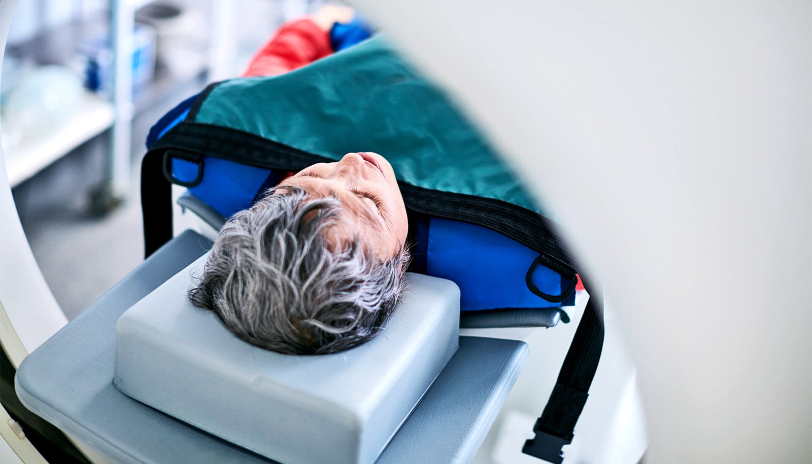
Early detection could get ahead of dementia damage
May 19, 2019 • futurityLinks/images:
- https://www.futurity.org/psychosis-feedback-striatum-1760582-2/
- https://doi.org/10.1038/s41386-019-0455-z
- https://nbsubscribe.missouri.edu/news-releases/2019/0827-neurological-brain-markers-might-detect-risk-for-psychotic-disorders/?utm_campaign=347932_08-27-19%20Kerns%20Psychological%20Disorders&utm_medium=email&utm_source=email&dm_i=42N5,7GGS,2H9PN8,S33F,1
- https://www.futurity.org/psychotic-disorders-risk-brain-mris-2144242/
- https://www.futurity.org