(Credit: Getty Images )
Technique suggests less invasive way to test fetal genes
A new technique that isolates trophoblast cells—placental cells that carry the complete fetal genome—could offer an alternative to more invasive tests.
A simple method of isolating trophoblast cells from the placenta could lead to less invasive ways of diagnosing genetic disorders in fetuses, a new study shows.
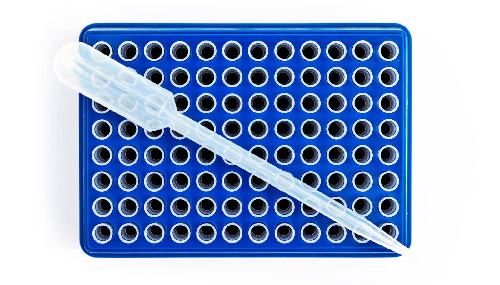
The technique isolates trophoblast cells—placental cells that carry the complete fetal genome—by taking advantage of their tendency to settle to the bottom of microwell plates. The researchers lay out a procedure for optimally isolating the cells, so they can pick them individually from the plate.
“This is the first study to use cell settling for enriching trophoblast cells from a heterogeneous cervical cell population,” the researchers write in Scientific Reports. “Ultimately, we provide a technique that is quick, inexpensive, minimizes cell loss, and results in retrieval of individual trophoblast cells.”
Currently, the only way to diagnose genetic disorders in developing fetuses involves retrieving trophoblasts through amniocentesis or chorionic villus sampling, both invasive procedures that carry a small risk of miscarriage.
Blood tests that look for fetal genetic material in the mother’s bloodstream can be useful screening tools, but scientists can’t use them for a definitive diagnosis. Further, screening is limited to whatever genetic material happens to turn up in the blood, which limits the range of disorders that can be screened.
Researchers know that trophoblasts are present in the cervical canal in the early stages of pregnancy, but the quantities are small, and isolating those cells from cervical cells and mucus is difficult.
For the new study, Christina Bailey-Hytholt, a PhD candidate in biomedical engineering at Brown University, and colleagues wanted to see if trophoblasts had any were any physical characteristics that might help in isolating them from cervical cells and other material.
They found that trophoblasts are smaller than cervical cells, differ in shape, and have relatively large nuclei. Those characteristics suggested that they may settle more quickly than cervical cells when they place cell mixtures on microwell plates.
Using polystyrene plates, the researchers found that the trophoblasts did indeed settle more quickly than cervical cells. The study showed that researchers achieved the maximum separation of cell types around four minutes after they put the cells on the plate. At that point, they could remove the cervical cells and mucus on top of the cells, leaving a large concentration of trophoblasts behind.
The technique increased the proportion of trophoblasts in samples by 700%, enabling individual trophoblasts to be picked out for genetic testing. The technique requires no specialized equipment beyond what any diagnostic lab would already have, the researchers say. And it takes only a few minutes to produce the cells necessary for genetic testing.
“There is a large need for biomedical engineering techniques toward advancing prenatal and women’s health,” Bailey-Hytholt says. “Our work is a step toward more non-invasive prenatal testing options.”
Additional coauthors are from Brown University and PerkinElmer, Inc. PerkinElmer, Inc. and the National Science Foundation supported the work.
Source: Brown University
The post Technique suggests less invasive way to test fetal genes appeared first on Futurity.
Share this article:
This article uses material from the Futurity article, and is licenced under a CC BY-SA 4.0 International License. Images, videos and audio are available under their respective licenses.
Related Articles:

Learning genetic risk can change how your body works
Dec. 12, 2018 • futurity
Pregnancy test false negatives are possible
April 25, 2019 • futurityLinks/images:
- https://www.futurity.org/placenta-brains-fetus-2007012/
- https://doi.org/10.1038/s41598-019-48346-3
- https://www.futurity.org/miscarriage-communicated-perspective-taking-1579452-2/
- https://www.brown.edu/news/2019-08-26/trophoblasts
- https://www.futurity.org/trophoblast-cells-placenta-genetic-testing-2151942-2/
- https://www.futurity.org