(Credit: Getty Images )
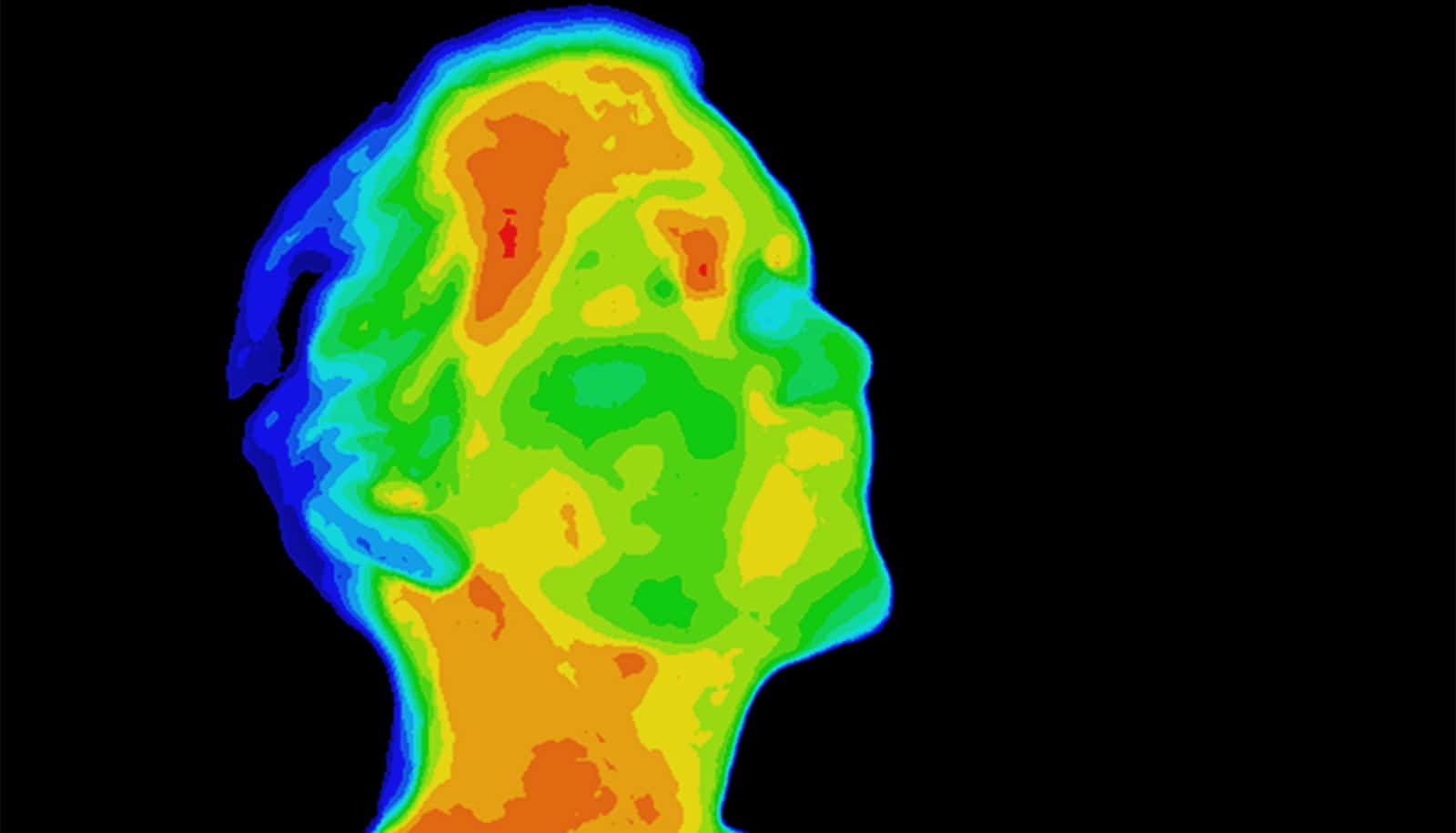
Infrared imaging shines a light on deep tumors
New infrared imaging technology can reveal tumors deep inside tissue with greater clarity than current methods, which could help guide treatment.

A new deep-tissue imaging technique can see beneath the skin of living subjects to illuminate buried tumors with unparalleled clarity, researchers report.
In a new study, the researchers demonstrate how their technique can help predict the response of cancer patients to immunotherapy and to track their progress following treatment.
“We call this infrared vision for non-invasively peering into biological tissues,” says study leader Hongjie Dai, professor in chemistry at Stanford University.
The technique relies on nanoparticles containing the element erbium, which belongs to a class of so-called rare-earth minerals chemists prize for their unique ability to glow in the infrared.
The team covered the nanoparticles in a chemically engineered coating that helps the particles dissolve in the bloodstream and also makes them less toxic and exit the body quicker. In addition, the coating provides anchoring points for molecules that that act like guided missiles to locate and attach to specific proteins on cells.
“It is like seeing while driving on a highway before and after the fog evaporates on a winter day…”
Under a low-powered LED light, the erbium particles bask targeted tissues or even individual cells in a bright infrared glow that can be seen deeper and with finer resolution than conventional imaging techniques, the researchers say.
“It is like seeing while driving on a highway before and after the fog evaporates on a winter day,” says study co-first author Yeteng Zhong, a postdoctoral researcher in Dai’s lab. “The combined imaging depth, molecular specificity and multiplicity, and spatial and temporal resolution are unattainable by previous techniques.”
In a video highlighting the power of their nanoprobes, the branching blood vessels of a living mouse’s brain glow with a teal light and shift as the mouse moves its head.
“Our approach allows for seeing into an intact mouse brain while conventional approaches see only the scalp,” says study co-first author Zhuoran Ma, a graduate student in Dai’s lab.
In the study, the researchers show their technique can identify tumors in mice carrying a protein that makes them vulnerable to cancer drugs that activate the body’s own immune system. This approach could provide a noninvasive way of identifying patients who would respond well to those drugs without needing to take a sample, or biopsy, of the tumor, as doctors currently need to do.
Similarly, erbium nanoparticles can monitor patients’ responses following cancer therapy to track whether they are responding to the drugs and if their tumors have shrunk.
As a demonstration, Dai and his colleagues combined their erbium nanoparticles with another infrared imaging technique to simultaneously image two molecular targets—cancer cells and T-cells—simultaneously. The result is a layered, multi-colored snapshot showing T-cells homing in on a tumor from elsewhere in the mouse’s body.
The researchers say their technique could also enable surgeons to more precisely excise tumors and aid biologists and medical researchers in studying fundamental processes within cells.
The research appears in the journal Nature Biotechnology. Additional researchers from Stanford and Beijing Jiaotong University also contributed to the work. Funding support for the research came from the National Institutes of Health.
Source: Stanford University
The post Infrared imaging shines a light on deep tumors appeared first on Futurity.
Share this article:
This article uses material from the Futurity article, and is licenced under a CC BY-SA 4.0 International License. Images, videos and audio are available under their respective licenses.
Related Articles:

Tweak amps up cancer immunotherapy by 77 fold
Oct. 1, 2021 • futurity
Invention uses nanoparticles to track chemo in the body
Sept. 26, 2019 • futurityLinks/images:
- https://www.futurity.org/nanoparticles-detect-cancer-1630312/
- https://www.futurity.org/breast-cancer-precise-lumpectomies-1894352/
- https://doi.org/10.1038/s41587-019-0262-4
- https://news.stanford.edu/2019/10/03/infrared-vision-immunotherapy/
- https://www.futurity.org/infrared-imaging-technology-cancer-2181722/
- https://www.futurity.org