Eye_orbit_anatomy_anterior2.jpg

Summary
| Description |
English:
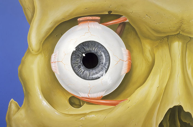
Normal anatomy of the human eye and orbit, anterior view.
Français :
Anatomie normale de l’œil humain et de l'orbite, en vue antérieure.
|
| Date | |
| Source | Patrick J. Lynch, medical illustrator |
| Author | Patrick J. Lynch, medical illustrator |
|
Permission
( Reusing this file ) |
Creative Commons Attribution 2.5 License 2006 |
| Other versions |
|

|
|
|
This image was selected as
picture of the day
on Wikimedia Commons for
10 June 2013
. It was captioned as follows:
English:
Normal anatomy of the human eye and orbit, anterior view.
Other languages:
English
:
Normal anatomy of the human eye and orbit, anterior view.
Français
:
Anatomie normale de l’œil humain et de l'orbite, en vue antérieure.
Nederlands
:
De normale anatomie van een menselijk
oog
en de
oogkas
(
orbinta
) van de voorzijde gezien.
한국어
:
앞쪽에서 바라본 사람의
눈
과 눈구멍의 해부학적 구조.
中文
:
人眼的解剖图
|
Patrick J. Lynch; illustrator; C. Carl Jaffe; MD; cardiologist Yale University Center for Advanced Instructional Media Medical Illustrations by Patrick Lynch, generated for multimedia teaching projects by the Yale University School of Medicine, Center for Advanced Instructional Media, 1987-2000. Patrick J. Lynch, http://patricklynch.net Creative Commons Attribution 2.5 License 2006; no usage restrictions except please preserve our creative credits: Patrick J. Lynch, medical illustrator; C. Carl Jaffe, MD, cardiologist https://creativecommons.org/licenses/by/2.5/ eye; anatomy; orbit; skull; rectus muscles; vision; human anatomy
-
You are free:
- to share – to copy, distribute and transmit the work
- to remix – to adapt the work
-
Under the following conditions:
- attribution – You must give appropriate credit, provide a link to the license, and indicate if changes were made. You may do so in any reasonable manner, but not in any way that suggests the licensor endorses you or your use.