Histology_bse.jpg
No higher resolution available.
| Description |
English:
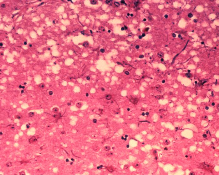
This micrograph of brain tissue reveals the cytoarchitectural histopathologic changes found in
bovine spongiform encephalopathy
. The presence of vacuoles, i.e. microscopic “holes” in the gray matter, gives the brain of BSE-affected cows a sponge-like appearance when tissue sections are examined in the lab.
Nederlands:
Deze microscopische opname toont hersenweefsel van een koe die aan BSE gestorven is. Tussen de hersencellen ziet men duidelijk verschillende
vacuoles
, die deze coupe (weefselsnede) een sponsachtig aanzicht geven.
Deutsch:
Das Bild zeigt die histopathologischen Veränderungen die bei einer Infektion mit BSE auftreten. Die Vakuolen, die in der grauen Substanz (substantia grisea) auftreten geben dem Bild ein schwamm-artiges Aussehen.
Français :
Cette coupe de tissu cérébral montre les modifications histopathologiques de l'organisation cellulaire lors d'une encéphalopathie spongiforme bovine. la présence de vacuoles, c'est-à-dire des "trous" microscopiques dans le tissu cérébral, donne au cerveau de vaches atteintes de l'ESB un aspect en éponge à l'examen des tissus en laboratoire.
|
| Date | |
| Source | Public Health Image Library, APHIS: http://www.aphis.usda.gov/lpa/issues/bse/bse_photogallery.html |
| Author | Dr. Al Jenny |
| Other versions | http://en.wikipedia.org/wiki/Image:Aphis.usda.gov_BSE_5.jpg |
|
|
This image or file is a work of a
United States Department of Agriculture
employee, taken or made as part of that person's official duties. As a
work
of the
U.S. federal government
, the image is in the
public domain
.
|

|