NMJ.jpg
No higher resolution available.
Summary
| Description |
English:
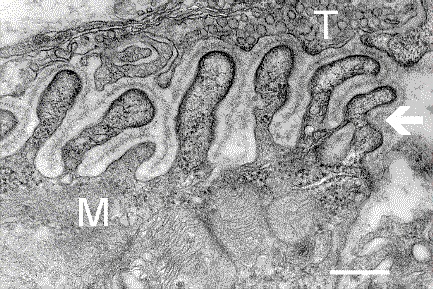
Electron micrograph
showing a cross-section through the
neuromuscular junction
. T is the
axon terminal
, M is the
muscle fiber
. The arrow shows junctional folds with
basal lamina
.
Postsynaptic densities
are visible on the tips between the folds. The scale is 0.3 µm.
|
| Date | Originally uploaded to en.wikipedia on 10 March 2006. |
| Source | Synapse Web at the National Institute of Mental Health, National Institutes of Health ; originally from en.wikipedia ; description page is/was here . |
| Author | National Institute of Mental Health ; originally uploaded by Nrets at en.wikipedia. |
Licensing
|
|
This image is a work of the
National Institutes of Health
, part of the
United States Department of Health and Human Services
, taken or made as part of an employee's official duties. As a
work
of the
U.S. federal government
, the image is in the
public domain
.

|
|
| This file has been identified as being free of known restrictions under copyright law, including all related and neighboring rights. | ||
Original upload log
(All user names refer to en.wikipedia)
- 2006-03-10 20:07 Nrets 433×289×8 (97758 bytes) Electron micrograph showing a cross section through the neuromuscular junction. T is the axon terminal, M is the muscle fiber. The arrow shows junctional folds with basal lamina. Postsynaptic densities are visible on the tips between the folds. Scale is 0