Panoramic_radiograph_of_historic_dental_implants.jpg
Size of this preview:
800 × 536 pixels
.
Other resolutions:
320 × 214 pixels
|
640 × 429 pixels
|
1,024 × 686 pixels
|
1,350 × 904 pixels
.
Summary
| Description |
English:
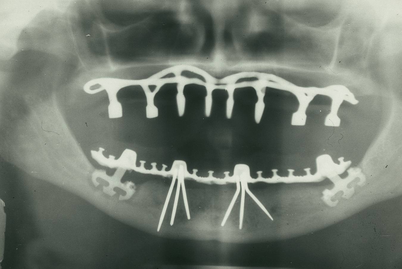
The x-ray controlling from 1976/77 shows an subperiosteal implant (according Cherchéve) in the maxilla. Two implant tripods (according Pruin) in the lower canine region and two stabilized blade Implants (according Heinrich) in the molar region.
Deutsch:
Die Röntgenaufnahme von 1976/77 zeigt ein subperiostales Implantat im Oberkiefer (Methode Cherchéve). In der unteren Eckzahnregion zwei Dreibeine aus Tantalnadeln (nach Pruin) sowie zwei stabilisierte Klingenimplantate (nach Heinrich). UK rechts Transluzenz beachten.
|
| Date | |
| Source | Own work |
| Author | Dentistxxx |
Licensing
I, the copyright holder of this work, hereby publish it under the following license:
This file is licensed under the
Creative Commons
Attribution-Share Alike 3.0 Unported
license.
-
You are free:
- to share – to copy, distribute and transmit the work
- to remix – to adapt the work
-
Under the following conditions:
- attribution – You must give appropriate credit, provide a link to the license, and indicate if changes were made. You may do so in any reasonable manner, but not in any way that suggests the licensor endorses you or your use.
- share alike – If you remix, transform, or build upon the material, you must distribute your contributions under the same or compatible license as the original.