Retina-diagram.svg

Size of this PNG preview of this SVG file:
743 × 331 pixels
.
Other resolutions:
320 × 143 pixels
|
640 × 285 pixels
|
1,024 × 456 pixels
|
1,280 × 570 pixels
|
2,560 × 1,140 pixels
.
| Description |
Deutsch:
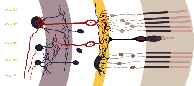
Axialer Aufbau der Retina (aus Cajal, 1911). (Cajal, 1991): S. R. Y. CAJAL, Histologie Du Système Nerveux de lHomme et Des Vertébrés, Maloine, Paris, 1911
Nervenzelltypen der Netzhaut schematisch, Licht fällt von links ein, weiß unterlegt die zellkernreichen Schichten v. l. n. r.: weiß: Ganglienzellen und ihre Axone, grau: Innere plexiforme Schicht, weiß: Amakrine Zellen, Bipolare, Horizontalzellen, gelb: Äußere plexiforme Schicht, weiß: Fotorezeptoren, hellbraun: Fotorezeptoren Außensegmente
English:
Axial organization of the retina (from Cajal, 1911). (Cajal, 1991): S. R. Y. CAJAL, Histologie Du Système Nerveux de lHomme et Des Vertébrés, Maloine, Paris, 1911
|
| Date | chris 論 12:06, 13 August 2009 (UTC) |
| Source | |
| Author |
|
|
Permission
( Reusing this file ) |
This file is licensed under the
Creative Commons
Attribution-Share Alike 3.0 Unported
license.
|