Validation_of_the_dye_diffusion_assay_performed_with_the_flattened_cochlear_preparation.png

Size of this preview:
287 × 599 pixels
.
Other resolutions:
115 × 240 pixels
|
230 × 480 pixels
|
979 × 2,044 pixels
.
Summary
| Description |
English:
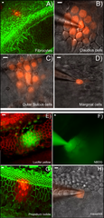
A–D: Dye diffusion patterns after PI was injected into a single cell in various locations in the cochlea. The type of the cells that was injected is given at lower right corner of each panel. E–F: Diffusion patterns of four different fluorescent dyes after injecting into a single Claudius cell. Name of the dye is given in the lower right corner of each panel. Panels B), C), D), F) & H) were photographed with unfixed fresh samples. Panels A), E), G) were results obtained from fixed samples after the experiments were done. They were labeled with fluorescent phalloidin (red in E, green in A&G) to outline the cell border. Scale bar on the top left of each panel represents approximately 100 µm.
|
| Date | |
| Source | PLOS ONE an open source peer reviewed journal- Gap Junction Mediated Intercellular Metabolite Transfer in the Cochlea Is Compromised in Connexin30 Null Mice ( [1] ) |
| Author | Qing Chang, Wenxue Tang, Shoeb Ahmad1, Binfei Zhou1, Xi Lin1 |
Licensing
This file is licensed under the
Creative Commons
Attribution 2.5 Generic
license.
-
You are free:
- to share – to copy, distribute and transmit the work
- to remix – to adapt the work
-
Under the following conditions:
- attribution – You must give appropriate credit, provide a link to the license, and indicate if changes were made. You may do so in any reasonable manner, but not in any way that suggests the licensor endorses you or your use.