
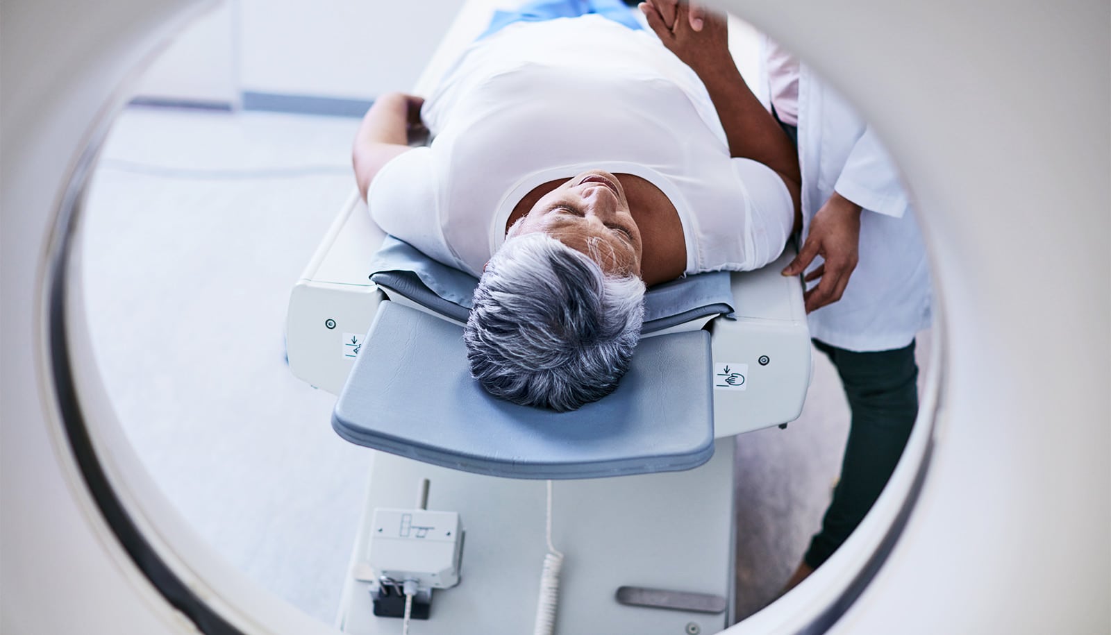
"...atrophied brain lesion volume represents a robust marker for predicting conversion from relapsing-remitting to secondary-progressive stages of MS."(Credit: Getty Images )
MRI marker better predicts progression of multiple sclerosis
Atrophied brain lesion volume is the only MRI marker to predict which cases of MS will progress to the more severe form of the disease, research finds.

Atrophied brain lesion volume is the only marker from MRI scans that can accurately predict which patients with multiple sclerosis will progress to the most severe form of the disease, a new study shows.
Secondary progressive MS, known as SPMS, typically appears 10 to 20 years after the initial onset and causes more physical and cognitive impairments.
Of the 1,314 patients in the 5-year study, more than 1,000 were women with an average age of 46.
The study in the journal Radiology builds on previous work that determined that atrophied brain lesion volume was a better indicator of disease progression than the appearance of lesions or whole brain atrophy, both of which researchers have long used as predictors of disease progression.
“This study corroborates initial reports from our group regarding using atrophied lesion volume as a potential MRI marker of disease progression in a large, population-based cohort of MS patients followed in clinical routine,” says Robert Zivadinov, professor of neurology in the Jacobs School at the University at Buffalo.
Brain lesion volume
As part of their routine care, patients with MS undergo MRI scans regularly, so that physicians can monitor new lesions or increased atrophy, which they see as indicators of more disease, or check for any reduction in lesions, generally seen as an indication that medications are working.
Getting Food and Drug Administration approval for a new MS drug requires, among other things, the ability of the new drug to reduce the number of brain lesions a patient has over 24 months.
But the new research demonstrates that atrophied brain lesion volume, which results from disintegration of lesions, and not creation of new lesions or progression of brain atrophy, more accurately signals progression of the disease.
“Neither changes in number and volume of lesions nor the development of whole brain or central brain atrophy showed any predictive power in demonstrating which patients would progress to secondary progressive MS, either from initial presentation of the disease, called clinically isolated syndrome, or the next stage, relapsing remitting MS,” Zivadinov says.
“The fact that atrophied lesion volume was the only measure that was predictive of conversion to progressive multiple sclerosis, and brain atrophy was not, is a major novel finding of this study.”
Signs of damage
Atrophied brain lesion volume can predict disease progression primarily because it reflects both inflammatory and neurodegenerative pathological processes, which together result in the disappearance of brain lesions into cerebrospinal fluid, Zivadinov says.
In previous work, Zivadinov and colleagues established an MRI marker, which measures brain lesions that cerebrospinal fluid has replaced. Lesions are signs of damage to the brain from physical trauma, a stroke, normal aging, or chronic disease. It is the disintegration of these lesions into cerebrospinal fluid that the researchers show is the main indicator of disease progression in MS.
“This study showed that atrophied brain lesion volume represents a robust marker for predicting conversion from relapsing-remitting to secondary-progressive stages of MS,” Zivadinov says.
The routine use of atrophied brain lesion volume as a marker for disease progression in MS depends on the completion of retrospective and prospective studies, some of which this team is already performing, on a large scale in clinical settings.
“Atrophied lesion volume can be measured with a pair of simple MRI scans,” Zivadinov says. “What has not been done yet is to test how visual or qualitative assessment compares to quantitative assessment performed in this study and previous studies we conducted.”
Researchers need a better understanding of the pathophysiological differences between those lesions that disappear compared to those that don’t. Future work will address this, Zivadinov says.
Source: University at Buffalo
The post MRI marker better predicts progression of multiple sclerosis appeared first on Futurity.
Share this article:
This article uses material from the Futurity article, and is licenced under a CC BY-SA 4.0 International License. Images, videos and audio are available under their respective licenses.
Related Articles:
New MRI method brings multiple sclerosis into sharper view
Feb. 12, 2024 • futurity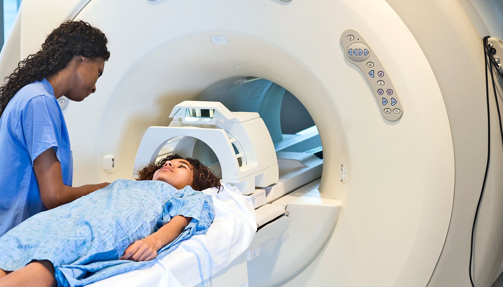
Multiple sclerosis often goes undetected in at-risk kids
March 20, 2023 • futurityLinks/images:
- https://doi.org/10.1148/radiol.2019190306
- https://www.futurity.org/multiple-sclerosis-lesions-brains-1774712-2/
- https://www.futurity.org/mri-metamaterial-2079022-2/
- http://www.buffalo.edu/news/releases/2019/002/034.html
- https://www.futurity.org/multiple-sclerosis-atrophied-brain-lesions-2168112-2/
- https://www.futurity.org