(Credit: Getty Images )
Imaging technique spots colorectal tumors with 100% accuracy
A new method that provides accurate, real-time, computer-aided diagnosis of colorectal cancer identified tumors with 100% accuracy in a new pilot study.

A new imaging technique in development provides accurate, real-time, computer-aided diagnosis of colorectal cancer, researchers say.
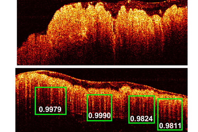
Using deep learning, a type of machine learning, researchers used the technique on more than 26,000 individual frames of imaging data from colorectal tissue samples to determine the method’s accuracy. Compared with pathology reports, the method identified tumors with 100% accuracy.
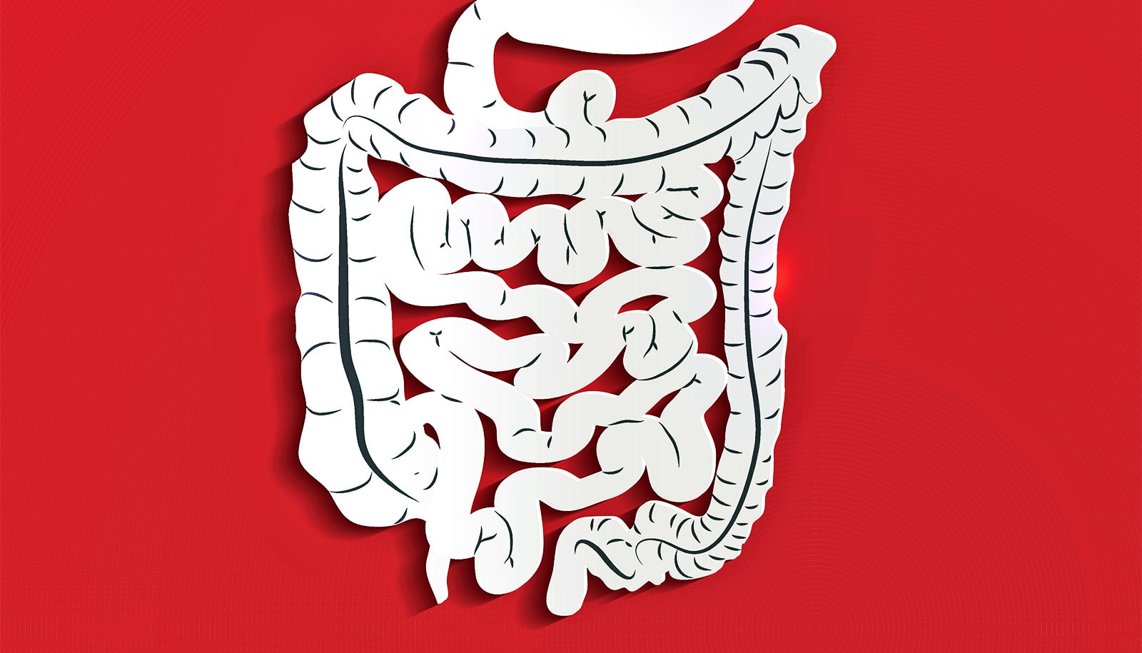
Colorectal cancer is the second most common type of cancer worldwide, with about 90% of cases occurring in people 50 or older. Arising from the inner surface, or muscosal layer, of the colon, cancerous cells can penetrate through the deeper layers of the colon and spread to other organs. Left untreated, the disease is fatal.
Currently doctors use flexible colonoscopy to perform colon cancer screening. The procedure involves visual inspection of the mucosal lining of the colon and rectum with a camera mounted on an endoscope. Doctors then biopsy abnormal appearing areas for analysis.
Although this is the current standard of care, it does have its shortcomings. First, the technique relies on visual detection, but small lesions are hard to detect with the naked eye, and often miss early malignancies. Second, visual endoscopy can only detect changes in the surface of the bowel wall, not in its deeper layers.
Imaging colorectal cancer
This is the first report using this type of imaging combined with machine learning to distinguish healthy colorectal tissue from precancerous polyps and cancerous tissue.
Researchers based the investigational technique on optical coherence tomography (OCT), an optical imaging technology used for two decades in ophthalmology to take images of the retina.
However, engineers have advanced the technology for other uses since it provides high spatial and depth resolution for up to 1- to 2-millimeter imaging depth. OCT detects the differences in the way health and diseased tissue refract light and is highly sensitive to precancerous and early cancer morphological changes.
When further developed, doctors could use the technique as a real-time, noninvasive imaging tool alongside traditional colonoscopy to assist with screening deeply seated precancerous polyps and early-stage colon cancers.
“We think this technology, combined with the colonoscopy endoscope, will be very helpful to surgeons in diagnosing colorectal cancer,” says senior author Quing Zhu, professor of biomedical engineering in the McKelvey School of Engineering and professor of radiology at the Mallinckrodt Institute of Radiology at Washington University in St. Louis.
“More research is necessary, but the idea is that when the surgeons use colonoscopy to examine the colon surface, this technology could be zoomed in locally to help make a more accurate diagnosis of deeper precancerous polyps and early-stage cancers versus normal tissue.”
From retinas to colons
Two years ago, lead author Yifeng Zeng, a biomedical engineering doctoral student, began using OCT as a research tool to image samples of colorectal tissue removed from patients at the School of Medicine. He observed that the healthy colorectal tissue had a pattern that looked similar to teeth. However, the precancerous and cancerous tissues rarely showed this pattern. Light attenuation of the healthy mucosa microstructures of the colorectal tissue caused the teeth pattern.
Zeng began working with another graduate student, Shiqi Xu, who earned a master’s in electrical engineering from McKelvey Engineering in 2019 and is co-first author of the paper, to train RetinaNet, a neural network model of the brain where neurons connect in complex patterns to process data, to recognize and learn the patterns in the tissue samples.
They trained and tested the network using about 26,000 OCT images acquired from 20 tumor areas, 16 benign areas, and six other abnormal areas in patient tissue samples. The diagnoses the system predicted compared with evaluation of the tissue specimens using standard histology.
Pathology residents Zahra Alipour and Heba Abdelal assisted with the comparison. The team found a sensitivity of 100% and a specificity of 99.7%.
“The unique part of our system is that we could detect a structural pattern within the image,” Zeng says. “Using OCT, we are imaging something that we can find a pattern across all normal tissues. Then we can use this pattern to classify abnormal and cancerous tissue for accurate diagnosis.”
The team is now developing a catheter that could be used simultaneously with the colonoscopy endoscope to analyze the teeth-like pattern on the surface of the colon tissue and to provide a score of probability of cancer from RetinaNet to the surgeons.
“Right now, we can obtain the feedback in 4 seconds,” Zeng says. “With further development of computation speed and the catheter, we can provide the feedback to surgeons in real-time,” Zeng says.
The research will appear in Theranostics.
The National Institutes of Health and the National Cancer Institute funded the work.
Source: Washington University in St. Louis
The post Imaging technique spots colorectal tumors with 100% accuracy appeared first on Futurity.
Share this article:
This article uses material from the Futurity article, and is licenced under a CC BY-SA 4.0 International License. Images, videos and audio are available under their respective licenses.
Related Articles:
Faster method spots anatomy to protect from radiation
Oct. 8, 2019 • futurity
MRI contrast agent detects early stage liver cancer
Feb. 10, 2020 • futurityLinks/images:
- https://www.futurity.org/colon-cancer-mutyh-protein-1790002/
- https://www.futurity.org/endoscope-cancer-chemotherapy-825692/
- https://www.futurity.org/can-a-new-vaccine-prevent-colon-cancer/
- https://www.futurity.org/robots-food-disabilities-caregivers-2006242-2/
- https://source.wustl.edu/2019/12/machine-learning-imaging-technique-may-boost-colon-cancer-diagnosis/
- https://www.futurity.org/colorectal-cancer-machine-learning-2227692-2/
- https://www.futurity.org