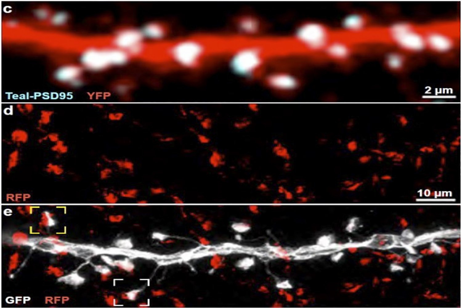
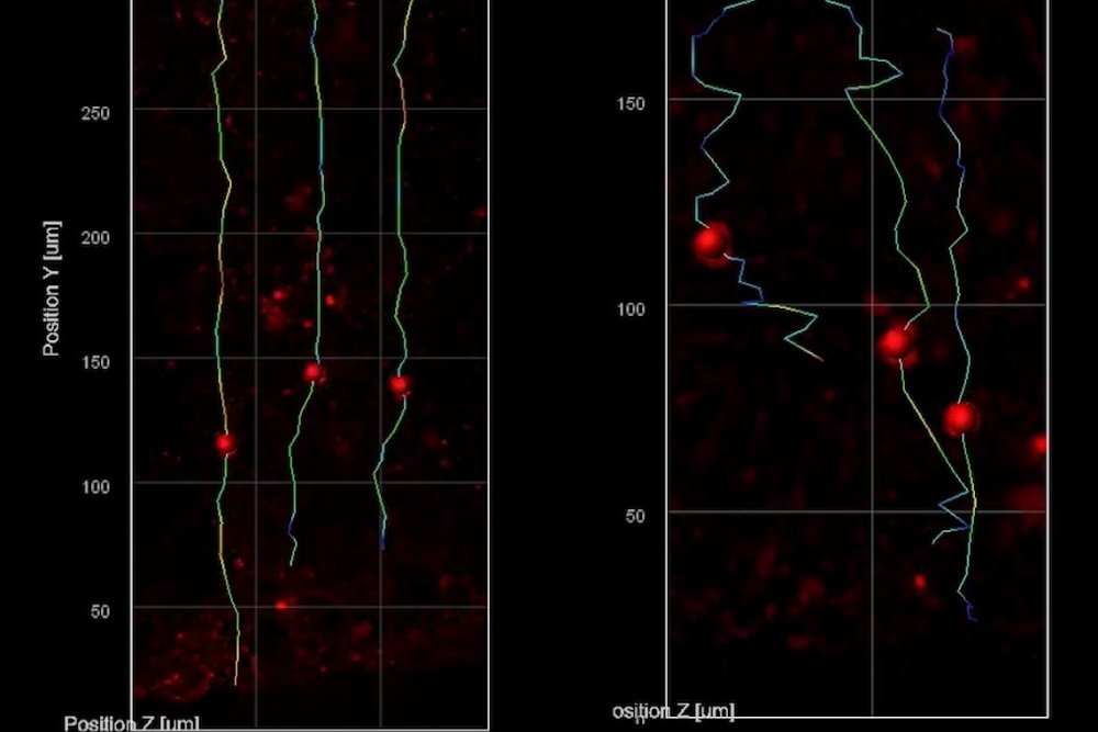
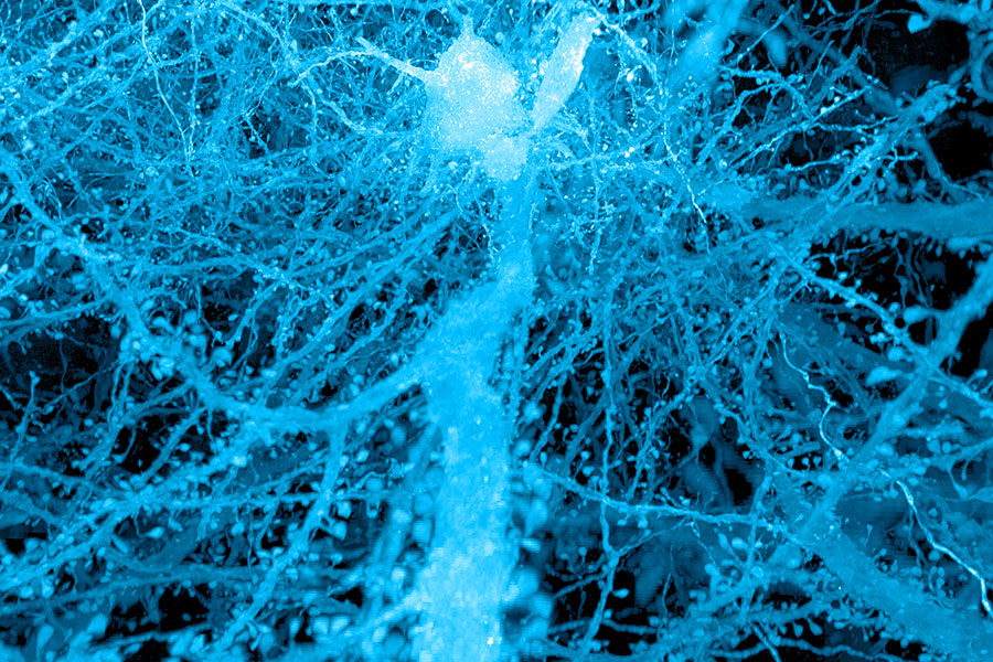
A brief history of expansion microscopy

Since an MIT team introduced expansion microscopy in 2015, the technique has powered the science behind kidney disease, plant seeds, the microbiome, Alzheimer’s, viruses, and more.

Jennifer Michalowski | McGovern Institute for Brain Research • mit
April 23, 2025 ~13 min

Seeing more in expansion microscopy

New methods light up lipid membranes and let researchers see sets of proteins inside cells with high resolution.

Jennifer Michalowski | McGovern Institute for Brain Research • mit
March 3, 2025 ~10 min


No detail too small

For Sarah Sterling, the new director of the Cryo-Electron Microscopy facility at MIT.nano, better planning and more communication leads to better science.

Nikole Fendler | Department of Biology • mit
Sept. 6, 2024 ~7 min

Elly Nedivi receives 2023 Kreig Cortical Kudos Discoverer Award

The neuroscientist is recognized for her ongoing work to understand molecular and cellular mechanisms that enable the brain to adapt to experience.

David Orenstein | The Picower Institute for Learning and Memory • mit
Nov. 29, 2023 ~3 min

A new way to see the activity inside a living cell

Using fluorescent labels that switch on and off, MIT engineers can study how molecules in a cell interact to control the cell’s behavior.

Anne Trafton | MIT News • mit
Nov. 28, 2023 ~8 min

Using AI to optimize for rapid neural imaging

MIT CSAIL researchers combine AI and electron microscopy to expedite detailed brain network mapping, aiming to enhance connectomics research and clinical pathology.

Rachel Gordon | MIT CSAIL • mit
Nov. 6, 2023 ~7 min

Exploring the nanoworld of biogenic gems

Project will develop new materials characterization tools and technologies to assign unique identifiers to individual pearls.

MIT.nano • mit
March 20, 2023 ~4 min

Sparse, small, but diverse neural connections help make perception reliable, efficient

First detailed mapping and modeling of thalamus inputs onto visual cortex neurons show brain leverages “wisdom of the crowd” to process sensory information.

David Orenstein | Picower Institute for Learning and Memory • mit
Feb. 2, 2023 ~9 min


Advanced imaging reveals mired migration of neurons in Rett syndrome lab models

Using organoids to model early development, researchers used an emerging microscopy technology to see that new neurons struggled to reach their developmental destination.

David Orenstein | Picower Institute for Learning and Memory • mit
Aug. 3, 2022 ~7 min

MIT.nano receives grant to acquire focused ion beam scanning electron microscope

National Science Foundation award will allow the VELION FIB-SEM to become a permanent instrument in MIT.nano’s characterization facility.

MIT.nano • mit
Feb. 9, 2022 ~4 min

/

4