Birefringence_microscopy_of_pseudogout,_annotated.jpg
Size of this preview:
662 × 600 pixels
.
Other resolutions:
265 × 240 pixels
|
530 × 480 pixels
|
848 × 768 pixels
|
1,130 × 1,024 pixels
|
1,745 × 1,581 pixels
.
Summary
| Description |
English:
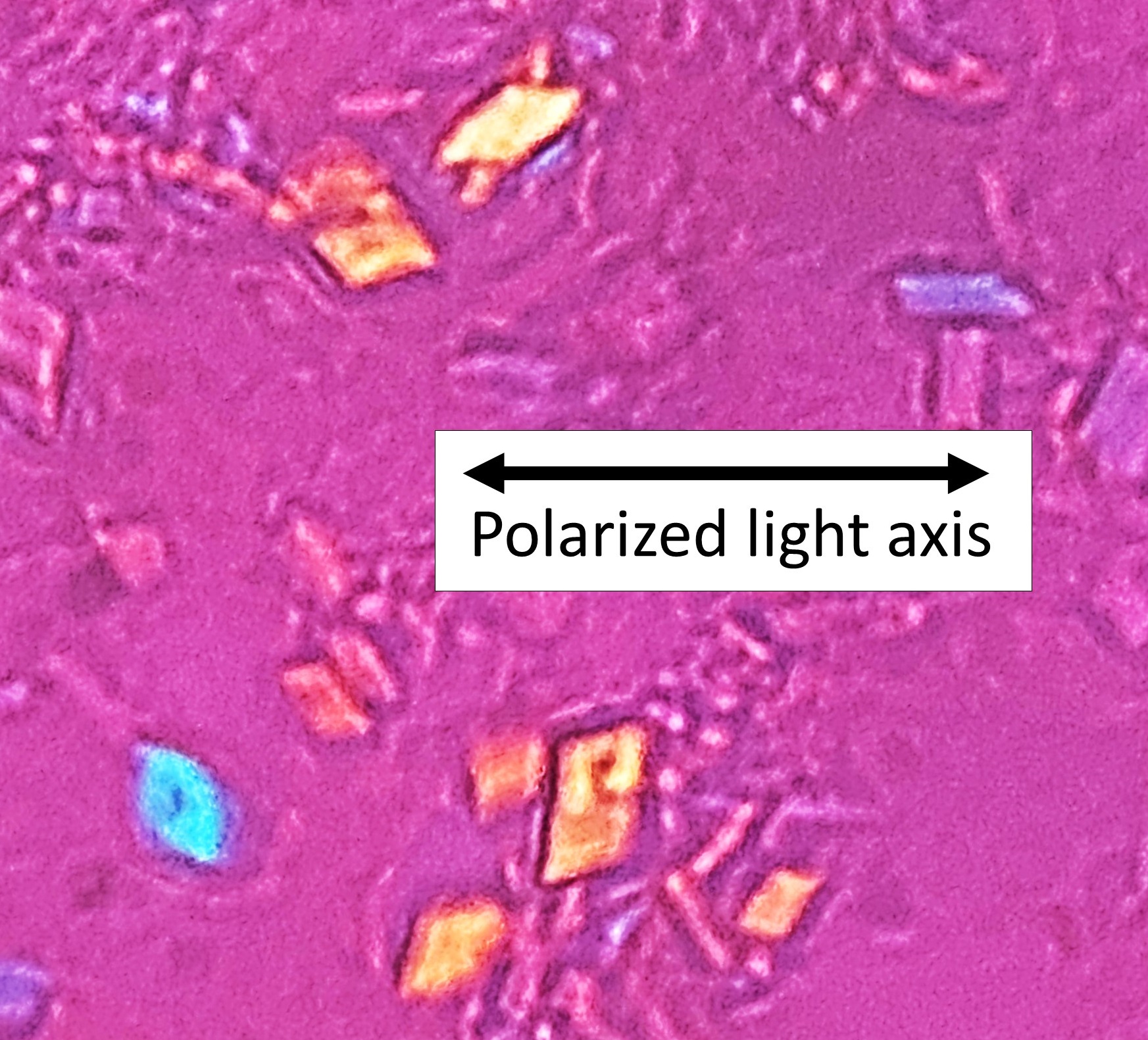
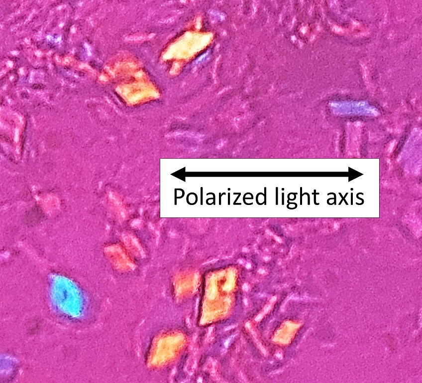
Microscopy with polarized light of tissue by a metatarsal joint, showing crystals whereof some (one annotated) have rhomboid shape and weak positive nirefringence, consistent with
calcium pyrophosphate dihydrate crystal deposition disease
(pseudogout).
|
| Date | |
| Source | Own work |
| Author |
.jpg)
- Reusing images - Conflicts of interest: None Consent note : Consent from the patient or patient's relatives is regarded as redundant, because of absence of identifiable features ( List of HIPAA identifiers ) in the media and case information ( See also HIPAA case reports guidance ). |
| Other versions |

|
| Camera location |
|
View this and other nearby images on: OpenStreetMap |
|
|---|
Licensing
|
|
This file is made available under the Creative Commons CC0 1.0 Universal Public Domain Dedication . |
|
The person who associated a work with this deed has dedicated the work to the
public domain
by waiving all of their rights to the work worldwide under copyright law, including all related and neighboring rights, to the extent allowed by law. You can copy, modify, distribute and perform the work, even for commercial purposes, all without asking permission.
|