Chlamydomonas_TEM_09.jpg

Size of this preview:
751 × 600 pixels
.
Other resolutions:
301 × 240 pixels
|
601 × 480 pixels
|
961 × 768 pixels
|
1,280 × 1,023 pixels
|
1,800 × 1,438 pixels
.
| Description |
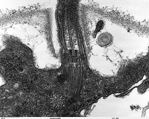
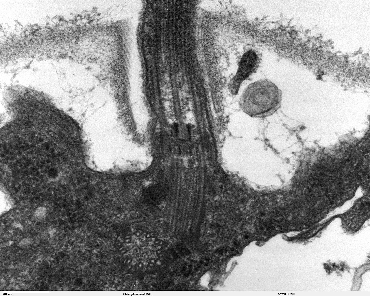
Transmission electron microscope image, showing an example of green algae (Chlorophyta).
Chlamydomanas reinhardtii is a unicellular flagellate used as a model system in molecular genetics work and flagellar motility studies. This image is a longitudinal section through the flagella area. In the cell apex is the basal body that is the anchoring site for a flagella. Basal bodies originate from and have a substructure similar to that of centrioles, with nine peripheral microtubule triplets(see structure at bottom center of image). The two inner microtubules of each triplet in a basal body become the two outer doublets in the flagella. This image also shows the transition region, with its fibers of the stellate structure. The top of the image shows the flagella passing through the cell wall. |
| Date | |
| Source | Source and public domain notice at http://remf.dartmouth.edu/imagesindex.html |
| Author | Dartmouth Electron Microscope Facility, Dartmouth College |
|
Permission
( Reusing this file ) |
Released into the public domain |
|
|
This work has been released into the
public domain
by its author,
Dartmouth Electron Microscope Facility, Dartmouth College
. This applies worldwide.
In some countries this may not be legally possible; if so: Dartmouth Electron Microscope Facility, Dartmouth College grants anyone the right to use this work for any purpose , without any conditions, unless such conditions are required by law.
|