Craniopharyngioma1.jpg

Size of this preview:
523 × 600 pixels
.
Other resolutions:
209 × 240 pixels
|
419 × 480 pixels
|
988 × 1,133 pixels
.
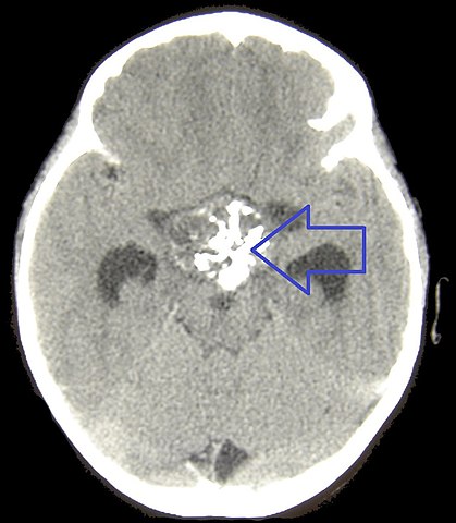
| Description | CT of a craniopharyngioma. This unenhanced CT shows a calcified cystic structure in the supra sellar region, together with hydrocephalus |
| Date | 6 August 2007 (upload date) |
| Source | Matthew R Garnett, Stéphanie Puget, Jacques Grill, Christian Sainte-Rose. Craniopharyngioma. Orphanet Journal of Rare Diseases. 2, 18. 2007. doi:10.1186/1750-1172-2-18 . PMID 17425791 |
| Author | see above |
This file is licensed under the
Creative Commons
Attribution 2.0 Generic
license.
-
You are free:
- to share – to copy, distribute and transmit the work
- to remix – to adapt the work
-
Under the following conditions:
- attribution – You must give appropriate credit, provide a link to the license, and indicate if changes were made. You may do so in any reasonable manner, but not in any way that suggests the licensor endorses you or your use.