Crc_met_to_node1.jpg

Size of this preview:
800 × 600 pixels
.
Other resolutions:
320 × 240 pixels
|
640 × 480 pixels
|
1,024 × 768 pixels
|
1,280 × 960 pixels
|
2,048 × 1,536 pixels
.
Summary
| Description |
English:
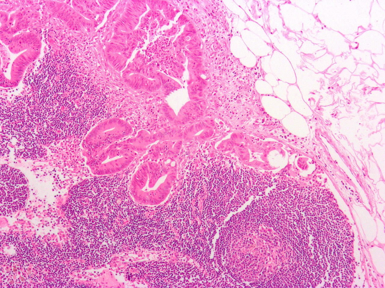
Micrograph
of a
colo
rectal
adenocarcinoma
metastasis
to a
lymph node
, also
lymph node metastasis
.
H&E stain
.
The cancer (forming glands) is seen at the centre-top. Adipose tissue is present on the upper right. See also
|
||
| Source | Own work | ||
| Author | Nephron | ||
|
Permission
( Reusing this file ) |
I, the copyright holder of this work, hereby publish it under the following licenses:
This file is licensed under the
Creative Commons
Attribution-Share Alike 3.0 Unported
license.
You may select the license of your choice.
|

| Annotations | This image is annotated: View the annotations at Commons |