Emphysema,_bullous,_subpleural_and_honeycomb_fibrosis_(4563270966).jpg

Size of this preview:
800 × 529 pixels
.
Other resolutions:
320 × 211 pixels
|
640 × 423 pixels
|
1,024 × 677 pixels
|
1,280 × 846 pixels
|
2,069 × 1,367 pixels
.
Summary
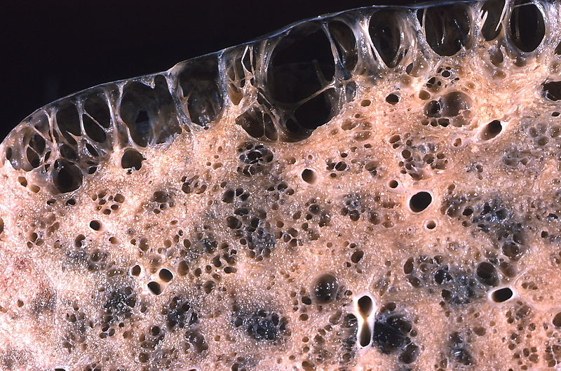
| Description | Large, prominent subpleural bullae . Spontaneous pneumothorax often results from rupture of such lesions. Honeycomb fibrosis is present in the subpleural lung parenchyma. In emphysema the spaces resulting from alveolar wall destruction have thin, delicate walls without intervening fibrosis and resemble the appearance of a spider web. In honeycomb fibrosis the abnormal spaces resulting both from alveolar wall destruction and dilatation of small airways are separated by broad bands of fibrous tissue thus resembling a bee's honeycomb. |
| Date | |
| Source |
Emphysema, bullous, subpleural and honeycomb fibrosis
|
| Author | Yale Rosen from USA |
Licensing
This file is licensed under the
Creative Commons
Attribution-Share Alike 2.0 Generic
license.
-
You are free:
- to share – to copy, distribute and transmit the work
- to remix – to adapt the work
-
Under the following conditions:
- attribution – You must give appropriate credit, provide a link to the license, and indicate if changes were made. You may do so in any reasonable manner, but not in any way that suggests the licensor endorses you or your use.
- share alike – If you remix, transform, or build upon the material, you must distribute your contributions under the same or compatible license as the original.
|
|
This image, originally posted to Flickr , was reviewed on 12 February 2014 by the administrator or reviewer File Upload Bot (Magnus Manske) , who confirmed that it was available on Flickr under the stated license on that date. |