Figure_1_BFDV_TEM_image.png

Size of this preview:
800 × 337 pixels
.
Other resolutions:
320 × 135 pixels
|
640 × 270 pixels
|
1,429 × 602 pixels
.
Summary
| Description |
English:
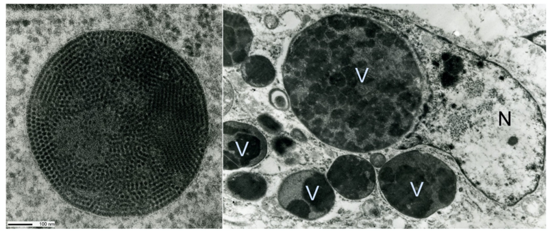
Transmission electron micrograph of BFDV infected cell on the right demonstrating how the nucleus (N) is relatively spared, with large crystalline arrays of mature virus particles preferentially forming intracytoplasmic inclusions (V) shown at higher magnification on the left (Sarker et al., 2016b).
|
| Date | |
| Source | Sarker, S., Terron, M.C., Khandokar, Y., Aragao, D., Hardy, J.M., Radjainia, M., Jimenez-Zaragoza, M., de Pablo, P.J., Coulibaly, F., Luque, D., Raidal, S.R., Forwood, J.K., 2016. Structural insights into the assembly and regulation of distinct viral capsid complexes. Nat Commun 7, 13014. |
| Author | Subir Sarker |
Licensing
This file is licensed under the
Creative Commons
Attribution 4.0 International
license.
-
You are free:
- to share – to copy, distribute and transmit the work
- to remix – to adapt the work
-
Under the following conditions:
- attribution – You must give appropriate credit, provide a link to the license, and indicate if changes were made. You may do so in any reasonable manner, but not in any way that suggests the licensor endorses you or your use.