Leaf_epidermis_w_scale.jpg

Size of this preview:
598 × 599 pixels
.
Other resolutions:
240 × 240 pixels
|
479 × 480 pixels
|
767 × 768 pixels
|
1,022 × 1,024 pixels
|
2,048 × 2,052 pixels
.
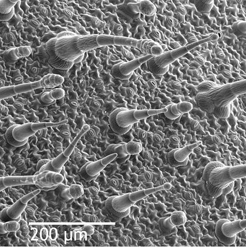
| Description | Scanning electron microscope image of Nicotiana alata upper leaf surface, showing tricomes and a few stomates. Instrument: ZEISS962 SEM. |
| Date | (UTC) |
| Source | |
| Author |
|
|
|
This is a
retouched picture
, which means that it has been digitally altered from its original version. Modifications:
Added scale, more contrast
. The original can be viewed here:
Leaf epidermis.jpg
:
|
|
|
This work has been released into the
public domain
by its author,
Louisa Howard
. This applies worldwide.
In some countries this may not be legally possible; if so: Louisa Howard grants anyone the right to use this work for any purpose , without any conditions, unless such conditions are required by law.
|
Original upload log
This image is a derivative work of the following images:
-
File:Leaf_epidermis.jpg
licensed with PD-author
- 2008-06-21T18:26:19Z Mangostar 2048x2073 (3038992 Bytes) {{Information |Description=Scanning electron microscope image of Nicotiana alata upper leaf surface, showing tricomes and a few stomates. Instrument: ZEISS962 SEM. |Source=http://remf.dartmouth.edu/images/NicotianaLeafSEM/nic
Uploaded with derivativeFX