Pericarditis.jpg
Size of this preview:
465 × 599 pixels
.
Other resolutions:
186 × 240 pixels
|
475 × 612 pixels
.
Summary
| Description |
English:
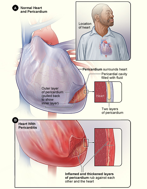
Figure A shows the location of the heart and a normal heart and pericardium (the sac surrounding the heart). The inset image is an enlarged cross-section of the pericardium that shows its two layers of tissue and the fluid between the layers. Figure B shows the heart with pericarditis. The inset image is an enlarged cross-section that shows the inflamed and thickened layers of the pericardium.
|
| Date | |
| Source | National Heart Lung and Blood Institute (NIH) |
| Author | National Heart Lung and Blood Institute (NIH) |
Licensing
|
|
This work is in the
public domain
in the United States because it is a
work prepared by an officer or employee of the United States Government as part of that person’s official duties
under the terms of
Title 17, Chapter 1, Section 105
of the
US Code
.
Note
: This only applies to original works of the Federal Government and not to the work of any individual
U.S. state
,
territory
, commonwealth, county, municipality, or any other subdivision. This template also does not apply to postage stamp designs published by the
United States Postal Service
since 1978
. (See §
313.6(C)(1)
of Compendium of U.S. Copyright Office Practices). It also does not apply to certain US coins; see
The US Mint Terms of Use
.
|

|
| This file has been identified as being free of known restrictions under copyright law, including all related and neighboring rights. | ||