SD-OCT_Corneal_Cross-Section.png
Size of this preview:
800 × 203 pixels
.
Other resolutions:
320 × 81 pixels
|
1,177 × 298 pixels
.
Summary
| Description |
English:
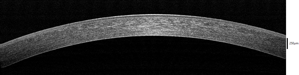
The healthy cornea of a 24 year old male (cross-section view). Corneal epithelium, stroma, and endothelium can be clearly seen.
This image is released to Wikimedia with patient consent. Imaged
in-vivo
with an Optovue iVue Spectral Domain Optical Coherence Tomographer (SD-OCT) at the office of Drs. Harry Wiessner, Steven Davis, Daniel Wiessner, and Eric Wiessner in Walla Walla, WA, USA.
|
| Date | |
| Source | Own work |
| Author | Wies6014 |
Licensing
I, the copyright holder of this work, hereby publish it under the following license:
This file is licensed under the
Creative Commons
Attribution-Share Alike 4.0 International
license.
-
You are free:
- to share – to copy, distribute and transmit the work
- to remix – to adapt the work
-
Under the following conditions:
- attribution – You must give appropriate credit, provide a link to the license, and indicate if changes were made. You may do so in any reasonable manner, but not in any way that suggests the licensor endorses you or your use.
- share alike – If you remix, transform, or build upon the material, you must distribute your contributions under the same or compatible license as the original.