Scanning_SQUID_microscopy.jpg
Size of this preview:
584 × 600 pixels
.
Other resolutions:
234 × 240 pixels
|
468 × 480 pixels
|
748 × 768 pixels
|
997 × 1,024 pixels
|
1,581 × 1,623 pixels
.
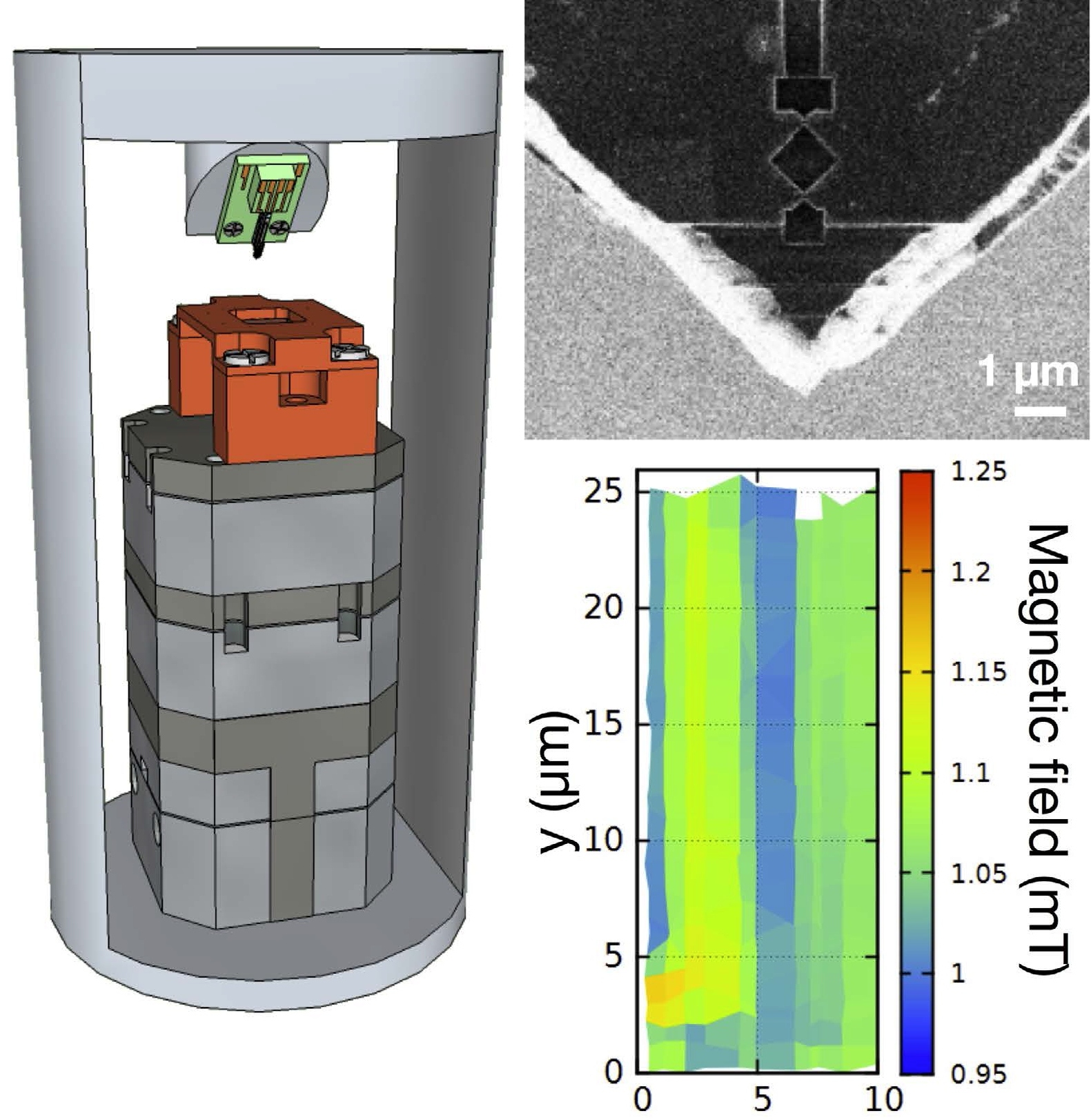
| Description | Left: Schematic of a scanning SQUID microscope in a 4 He refrigerator. Green holder for the SQUID probe is attached to a quartz tuning fork. Bottom part is a piezoelectric sample stage. Right: electron micrograph of a SQUID probe and a test image of Nb/Au strips recorded with it. |
| Date | |
| Source | http://www.nature.com/articles/srep15097 |
| Author | Yusuke Shibata, Shintaro Nomura, Hiromi Kashiwaya, Satoshi Kashiwaya, Ryosuke Ishiguro & Hideaki Takayanagi |
|
Permission
( Reusing this file ) |
This file is licensed under the
Creative Commons
Attribution 4.0 International
license.
|