TRNA-Phe_yeast_1ehz.png

Size of this preview:
606 × 599 pixels
.
Other resolutions:
243 × 240 pixels
|
485 × 480 pixels
|
777 × 768 pixels
|
1,174 × 1,161 pixels
.
Summary
| Description |
English:
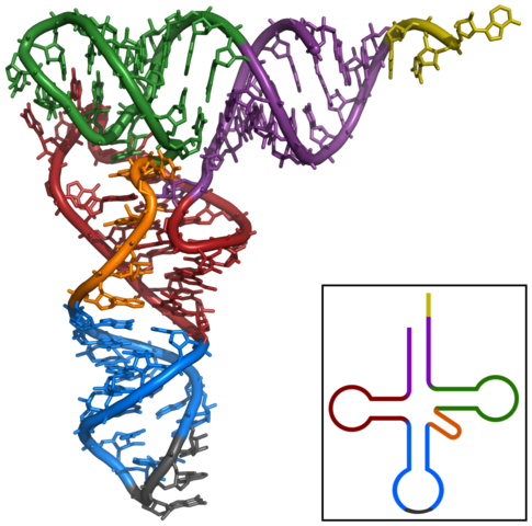
X-ray structure of the tRNA
Phe
from yeast. Data was obtained by
PDB
:
1ehz
and rendered with PyMOL.
Deutsch:
Struktur der tRNA
Phe
aus Hefe. Dem Bild liegen die Daten aus
PDB
:
1ehz
zu Grunde, es wurde mittels PyMOL gerendert.
Español:
Estructura de rayos X de la tRNA
Phe
de la levadura. Los datos se obtuvieron por AP: 1ehz y se procesaron con PyMOL.
|
| Date | |
| Source | Own work |
| Author | Yikrazuul |
Licensing
I, the copyright holder of this work, hereby publish it under the following license:
This file is licensed under the
Creative Commons
Attribution-Share Alike 3.0 Unported
license.
-
You are free:
- to share – to copy, distribute and transmit the work
- to remix – to adapt the work
-
Under the following conditions:
- attribution – You must give appropriate credit, provide a link to the license, and indicate if changes were made. You may do so in any reasonable manner, but not in any way that suggests the licensor endorses you or your use.
- share alike – If you remix, transform, or build upon the material, you must distribute your contributions under the same or compatible license as the original.