Tuberculosis-x-ray.jpg
No higher resolution available.
Summary
| Description |
English:
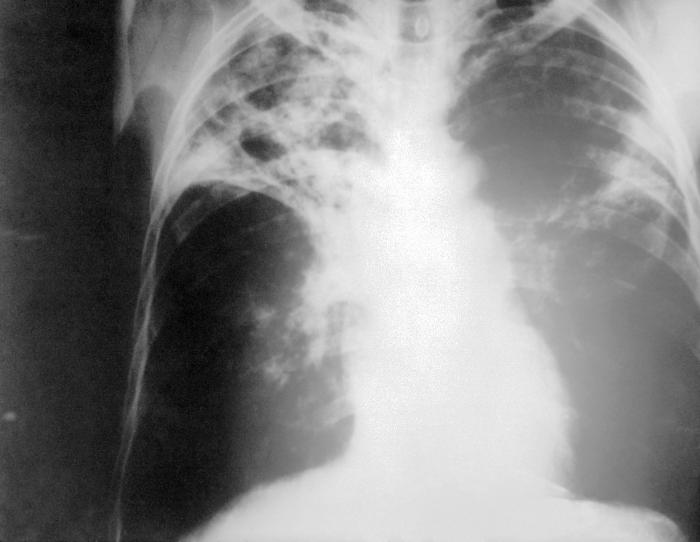
An anteroposterior X-ray of a patient diagnosed with advanced bilateral pulmonary tuberculosis. This AP X-ray of the chest reveals the presence of bilateral pulmonary infiltrate, and „caving formation“ present in the right apical region. The diagnosis is far-advanced tuberculosis.
Deutsch:
Eine Röntgenaufnahme im anterior-posterioren Strahlengang eines Patienten, bei dem eine beidseitige Lungentuberkulose festgestellt wurde. Diese Thorax-Aufnahme zeigt beidseitige Lungeninfiltrate und eine sogenannte „Kaverne“ im rechten Oberfeld. Sie entspricht der Diagnose einer fortgeschrittenen Lungentuberkulose.
|
||
| Date |
|
||
| Source |
|
||
| Author |
|
||
|
Permission
( Reusing this file ) |
PD-USGov-HHS-CDC
English:
None - This image is in the public domain and thus free of any copyright restrictions. As a matter of courtesy we request that the content provider be credited and notified in any public or private usage of this image.
|
Licensing
|
|
This image is a work of the
Centers for Disease Control and Prevention
, part of the
United States Department of Health and Human Services
, taken or made as part of an employee's official duties. As a work of the
U.S. federal government
, the image is in the
public domain
.
|
|
Original upload log
The original description page was
here
. All following user names refer to en.wikipedia.
- 2005-06-05 14:21 Rsabbatini 700×542×8 (32649 bytes) An anteroposterior [[X-ray]] of a patient diagnosed with advanced bilateral [[lung|pulmonary]] [[tuberculosis]]. This AP X-ray of the [[chest]] reveals the presence of bilateral pulmonary infiltrate, and “caving formation” present in the right apical