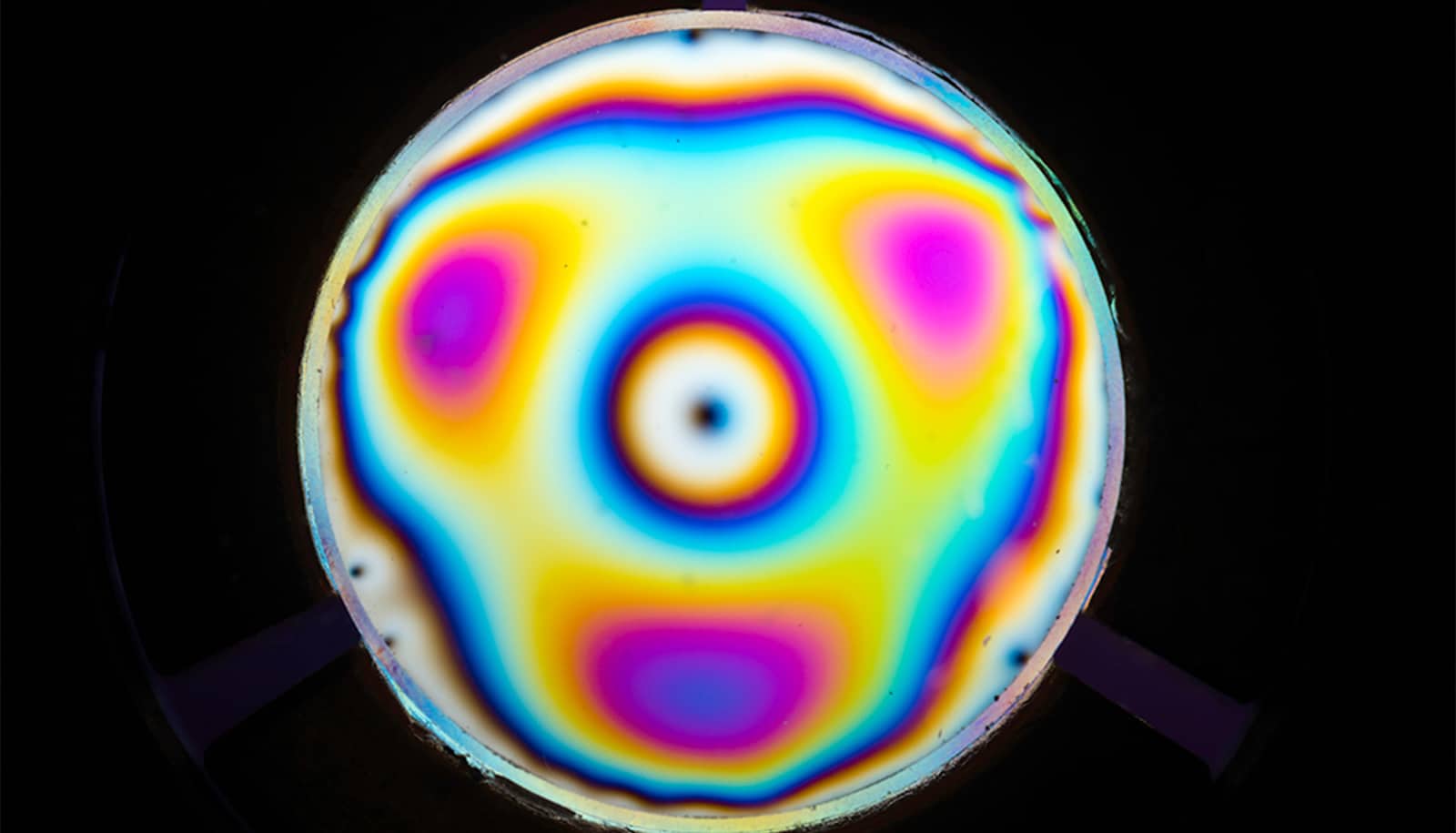
A new microscopy system that can can image individual molecules in 3D and capture the way they “wobble” uses a specially engineered glass plate. (Credit: J. Adam Fenster/U. Rochester)
Imaging tech captures molecule ‘wobble’ in 3D
Called CHIDO, Mexican slang for "cool," the new technology takes imaging single molecules into the third dimension.
Researchers have found a way to visualize individual molecules inside living cells in greater detail, showing their position and orientation in 3D, and even how they wobble and oscillate.
The work could shed invaluable insights into the biological processes involved, for example, when a cell and the proteins that regulate its functions react to the virus that causes COVID-19.
“When a protein changes shape, it exposes other atoms that enhance the biological process, so the change of shape of a protein has a huge effect on other processes inside the cell,” says Sophie Brasselet, director of the Fresnel Institute in France, who collaborated with Miguel Alonso and Thomas Brown, both professors of optics at the University of Rochester.
The researchers describe their new technology, nicknamed CHIDO (“Coordinate and Height super-resolution Imaging with Dithering and Orientation”), in a new paper in Nature Communications .
A new view in 3D
CHIDO is precise within “tens of nanometers in position and a few degrees of orientation” in determining the parameters of single molecules,” the researchers report.
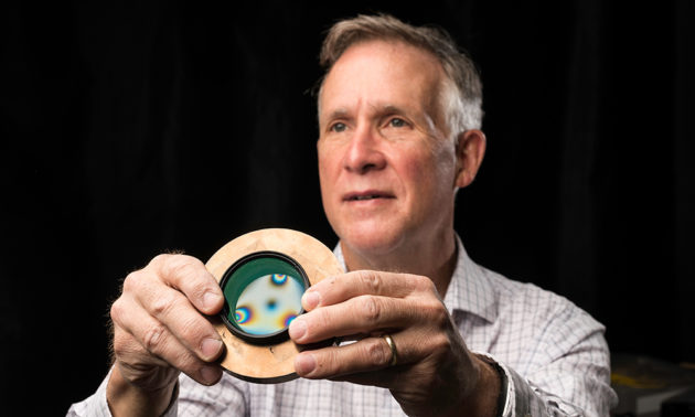
Using a glass plate subjected to uniform stress all around its periphery, the device can create and extrapolate wavelength oscillations and changes in polarization that occur when molecules are observed in a fluorescence microscope.
The new technology transforms the image of a single molecule into a distorted focal spot, the shape of which directly encodes more precise 3D information than previous measurement tools. In effect, CHIDO can produce beams that have every possible polarization state.
“This is one of the beauties of optics,” Brown says. “If you have a device that can create just about any polarization state, then you also have a device that can analyze just about any possible polarization state.”
The glass plate originated in Brown’s lab as part of his long interest in developing beams with unusual polarizations. Alonso, an expert on the theory of polarization, worked with Brown on ways to refine this “very simple but very elegant device” and expand its applications.
During a visit to Marseille, Alonso described the plate to Brasselet, an expert in novel instrumentation for fluorescence and nonlinear imaging. Brasselet immediately suggested its possible use in the microscopy techniques she was working on to image individual molecules.
“It’s been a very complementary team,” Brasselet says.
The origin of CHIDO
In 1873, Ernst Abbe stipulated that microscopes would never obtain better resolution than half the wavelength of light. That barrier stood until Nobel laureates Eric Betzig and William Moerner—with their single-molecule microscopy—and Stefan Hell—with his stimulated emission depletion microscopy—found ways to bypass it.
Six years ago, the Nobel committee awarded them the chemistry prize for finding ways to visualize the pathways of individual molecules inside living cells.
“Due to their achievements the optical microscope can now peer into the nanoworld,” the Nobel committee reported in 2014.
“What was missing in that Nobel Prize and the work in subsequent years was the ability to not only accurately know the location of a molecule, but to be able to characterize its direction and especially its motion in three dimensions,” Brown says.
In fact, the solution Brown, Alonso, and Brasselet now describe had its origins 20 years ago.
Starting in 1999, Brown and one of his PhD students, Kathleen Youngworth, began investigating unusual optical beams that displayed unusual patterns of optical polarization, the orientation of the optical wave. Some of the beams exhibited a spoke-like radial pattern with intriguing properties.
Youngworth demonstrated on a tabletop that, when tightly focused, the beams exhibited polarization components that pointed in almost any direction in three dimensions.
Alexis Spilman Vogt, another PhD candidate, then worked with Brown on creating the same effects by applying stress to the edges of a glass cylinder. Brown’s brother-in-law, Robert Sampson, a skilled tool and die specialist, was called upon to fabricate some samples and fit them in metal rings for use with a confocal microscope.
This involved heating both the glass and metal rings. “Metal expands at a faster rate when you heat it than glass does,” Brown says, “and so you could heat the glass and metal up very hot, insert the glass in the middle of the metal, and as it cools down the metal would shrink and create a tremendous force on the periphery of the glass.”
Sampson inadvertently applied more stress than called for with one of the plates. As soon as his brother-in-law handed it to him, Brown knew the plate had unusual qualities. The Rochester group introduced the term “stress engineered optic” to describe the elements and, as they learned more about both the physical behavior and the mathematics, they realized that the windows could be the path the solving entirely new problems in microscopy.
And that was the origin of what is now CHIDO, which, coincidently, happens to be Mexican slang for “cool.”
“At the time Alexis and I knew the stress-engineered glass was interesting, and would likely have useful applications; we just didn’t know at the time what they might be,” Brown says.
Now, thanks to his collaboration with Alonso and Brasselet, he hopes CHIDO will “catch the imagination” of other researchers in the field who can help further refine and apply the technology.
Support for the research came from the National Science Foundation, the Excellence Initiative of Aix-Marseille University, the European Union’s Horizon 2020 research and innovation program, and the CONACYT Doctoral Fellowship program.
Source: University of Rochester
The post Imaging tech captures molecule ‘wobble’ in 3D appeared first on Futurity .
Share this article:
This article uses material from the Futurity article, and is licenced under a CC BY-SA 4.0 International License. Images, videos and audio are available under their respective licenses.
Related Articles:
X-ray ‘movie’ captures molecular motion in real time
July 17, 2019 • futurity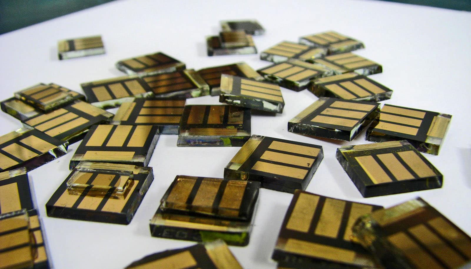
Team sees light make atoms ‘dance’ in perovskites
Feb. 14, 2023 • futurityLinks/images:
- https://www.futurity.org/sars-cov-2-virus-evolutionary-history-2411662/
- https://doi.org/10.1038/s41467-020-19064-6
- https://www.futurity.org/machine-learning-microscope-2217132/
- https://www.futurity.org/glass-phase-transition-physics-2327992/
- https://www.rochester.edu/newscenter/finally-a-way-to-see-molecules-wobble-458232/
- https://www.futurity.org/chido-visualizing-single-molecules-2459062/
- https://www.futurity.org